3D Printing
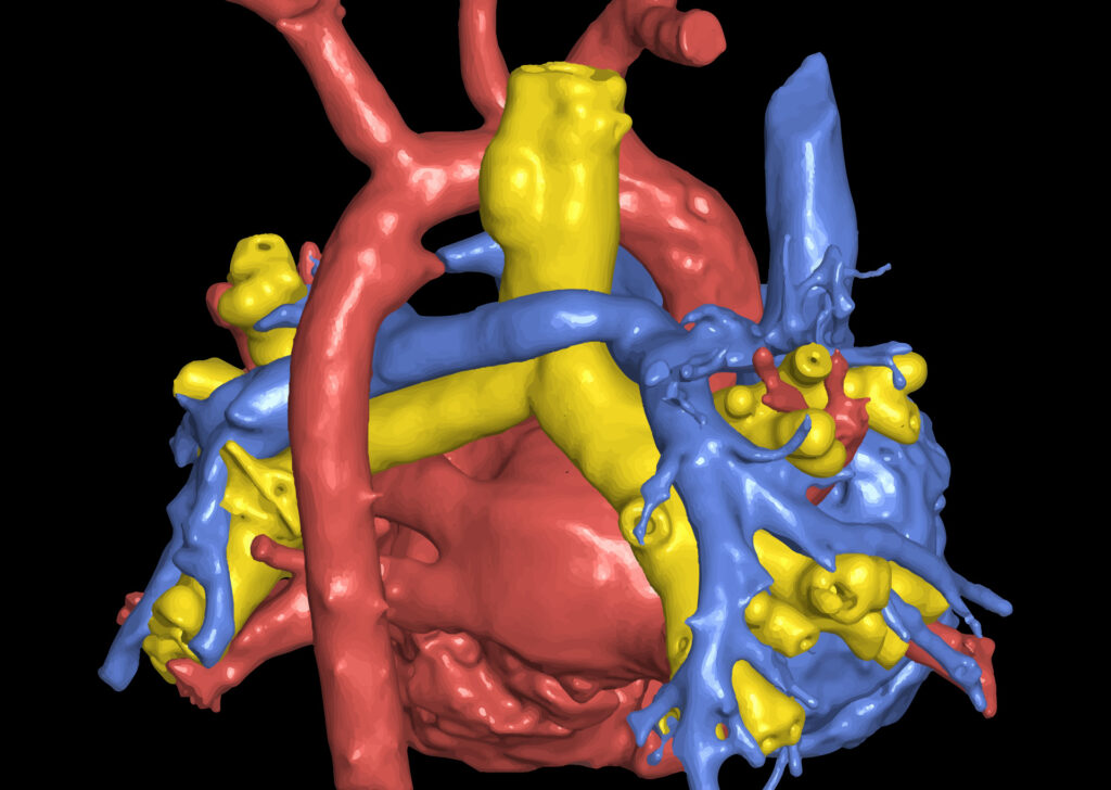
Converting digital medical images into 3D printed models is revolutionizing the healthcare industry. Vitrea software provides expert segmentation tools incorporated with the ability to export stereolithography (STL), OBJ (waveform object) and VRML (virtual reality modeling language) files for 3D modeling.
The 3D output anatomical model is not for diagnostic use.
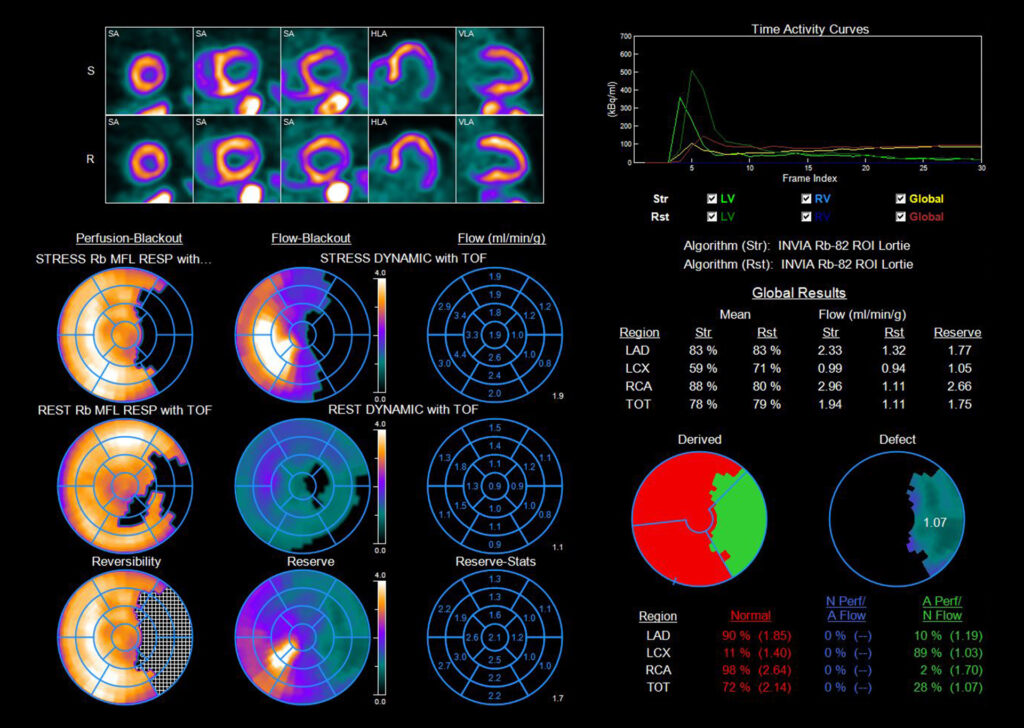
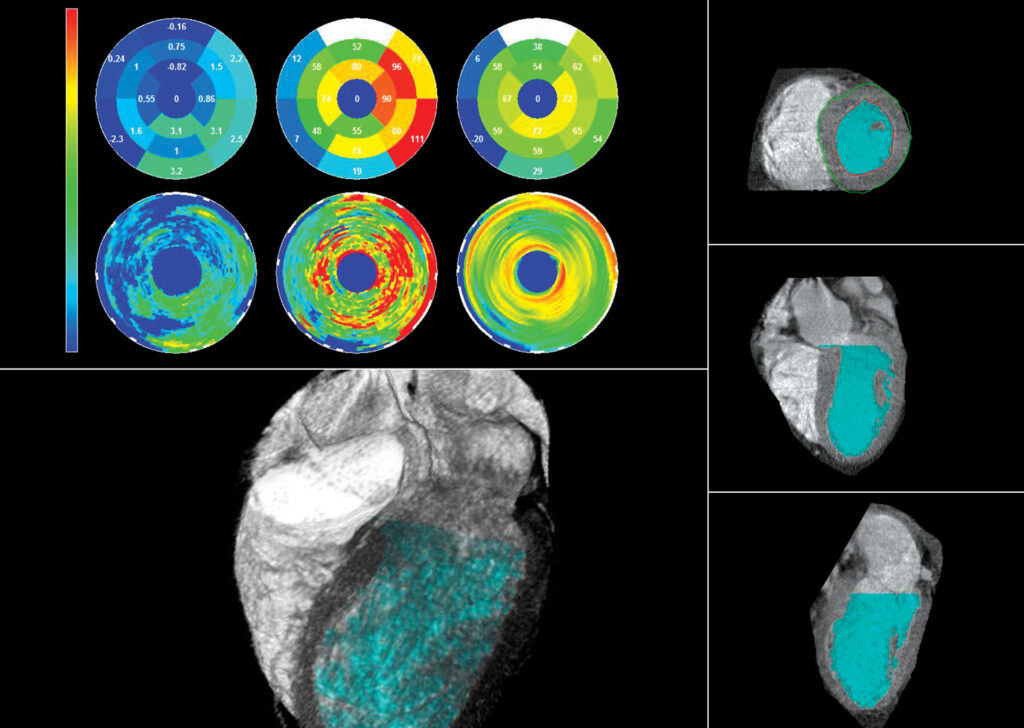
4DM powered by INVIA
4DM is integrated into Vitrea Advanced Visualization and is state-of-the-art software for cardiovascular quantification, image review and reporting of SPECT and PET patient studies.
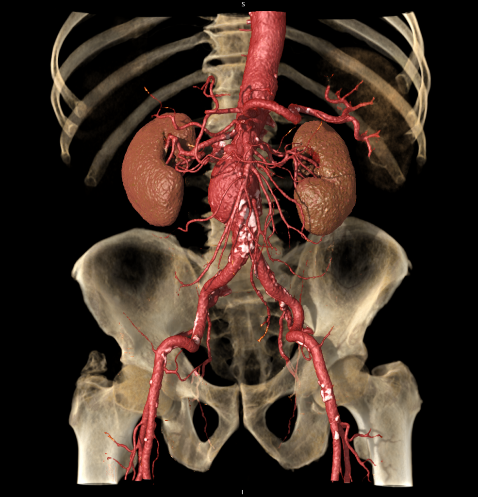
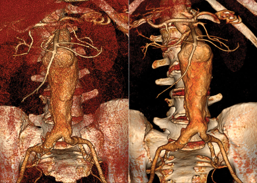
CT Aorta Analysis
CT Aorta Analysis enables users to visualize and evaluate the aorta vasculature.
CT Cardiac Analysis
CT Cardiac Analysis enables physicians to determine the presence and extent of coronary obstructive disease by displaying the extracted anatomy in a variety of views. The interface and automated tools help to efficiently analyze the coronary arteries.
CT Cardiac Functional Analysis
CT Cardiac Functional Analysis (CFA) utilizes CT images of the heart to assist cardiologists and radiologists in assessing cardiac function for the left ventricle.
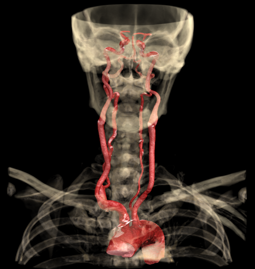
CT Carotid
CT Carotid provides tools to visualize and evaluate the carotid and vertebral vessels.
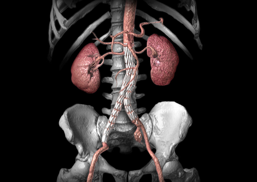
CT Endovascular Stent Planning (EVSP)
CT EVSP enables visualization and measurements of aortic vessels for evaluation, treatment and follow-up for aortic vascular disorders.
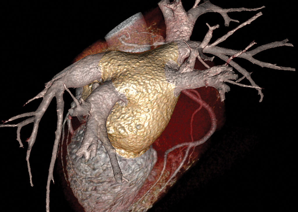
CT EP Planning
CT EP Planning enables analysis and assessment of the left atrium and pulmonary veins. The application provides optimized 2D and 3D views with tools for quantitative measurements and 3D model export capabilities.
CT Multi-Chamber CFA
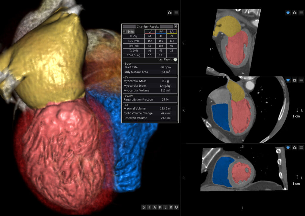
CT Multi-Chamber CFA utilizes CT images of the heart to assist cardiologists and radiologists in assessing cardiac function of the heart’s individual chambers.
CT Myocardial Perfusion
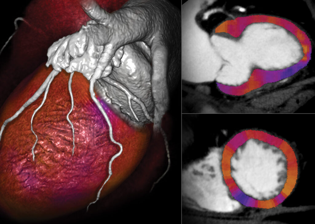
CT Myocardial Perfusion enables the visualization and analysis of perfusion deficits in the myocardium. Semi-automated segmentation and registration are available in a streamlined workflow.
CT Peripheral
CT Peripheral vessels can be post-processed in various ways. Auto Bone Removal and Vessel Grow provide a quick overview of the peripheral arteries. The Vessel Probe option segments and evaluates contrast-filled arteries. The software permits you to easily calculate arterial stenosis and plaque burden.
CT Renal
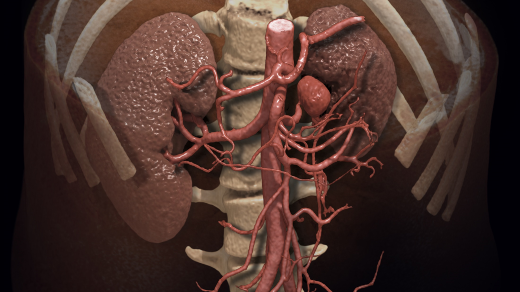
CT Renal enables the visualization of renal anatomy using CT angiography studies.
CT SUREPlaque™
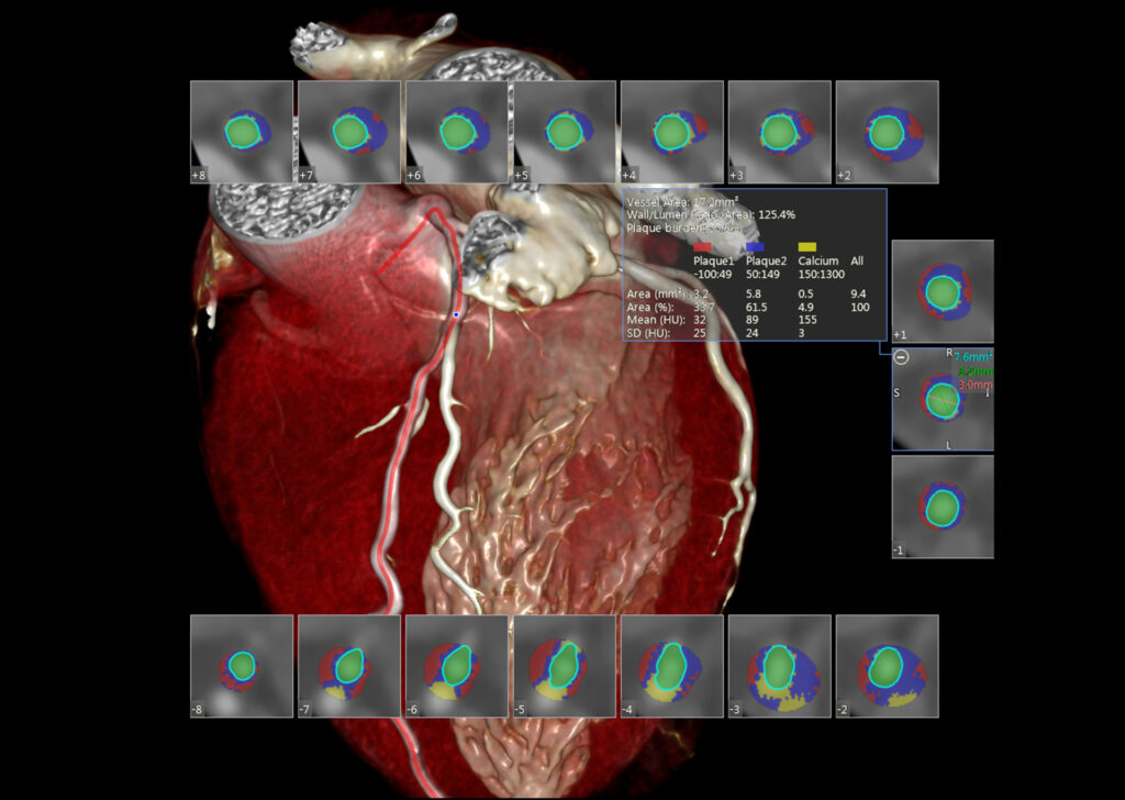
CT SUREPlaque provides the visualization and measurement of vessel walls and plaque characteristics in arterial vessels using color defined Hounsfield Unit (HU) ranges through a streamlined workflow.
CT Transcatheter Aortic Valve Replacement (TAVR) Planning
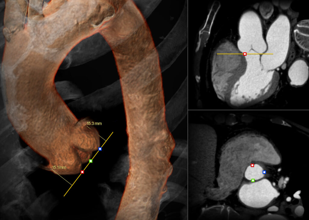
CT TAVR Planning assists with the assessment of the aortic valve and in pre-operative planning and post-operative evaluation of transcatheter aortic valve replacement procedures.
CT VScore™
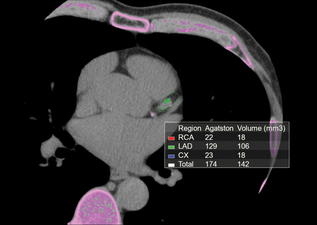
CT VScore is a calcium scoring application that provides the ability to visualize, measure and create a report of coronary calcification and calculate the calcium score using a non-contrast cardiac CT exam.
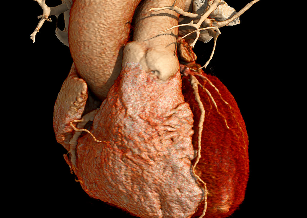
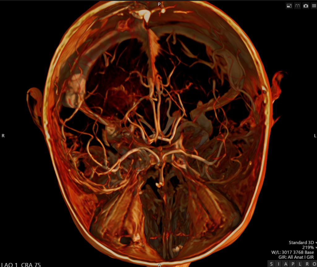
Global Illumination
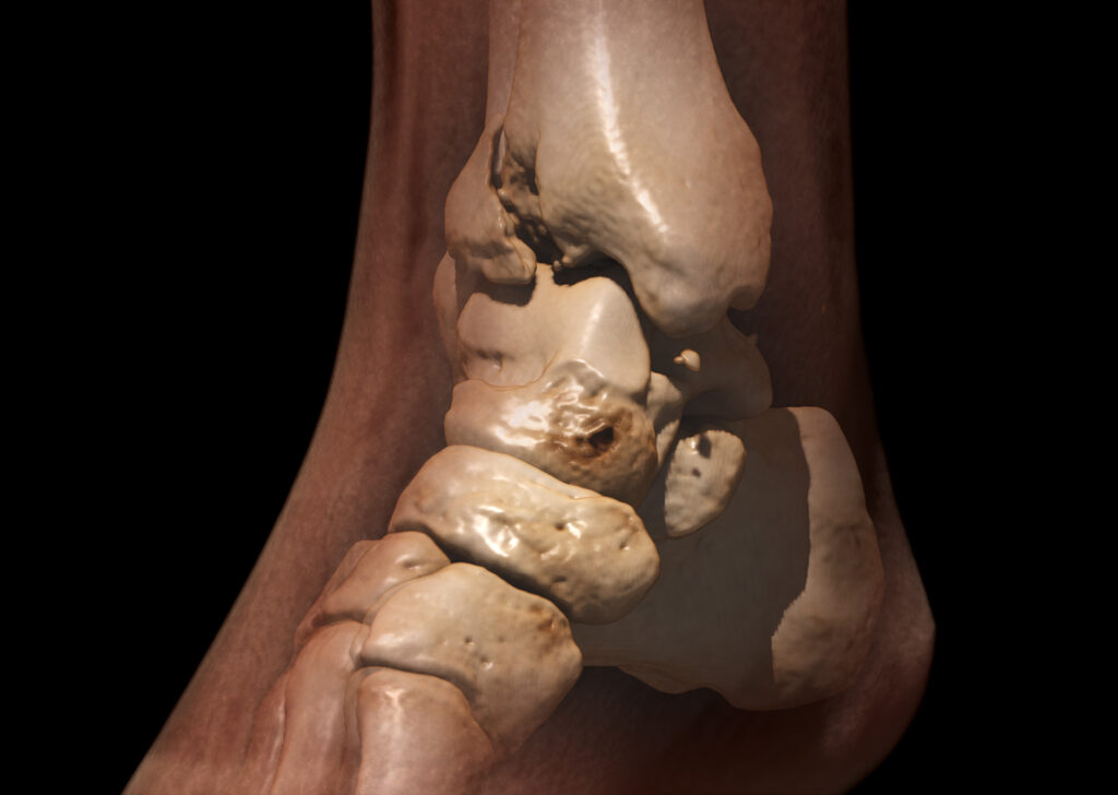
Global Illumination is an alternate 3D rendering technique to help provide a more photorealistic view of human anatomy. Users are able to acquire and share these images for communication and education.
Medis® Suite Cardiovascular MR (CVMR)
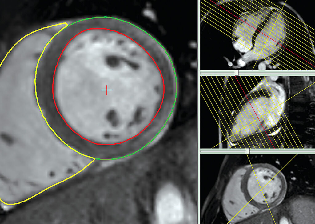
This suite allows for efficient and easy processing of Cardiovascular MR cases, with industry-leading QMass® and QFlow® analytical applications. 4D Flow allows visualization and quantification of 4D Flow datasets. QStrain allows LV, RV and Atrial strain analysis.
MR Vascular
The MR Vascular workflow evaluates vascular anatomy from MR angiography studies.
Vitrea Image Denoising
Image Denoising is a software-based, post-processing filter designed to be used in conjunction with original image data. It assists clinicians in the enhancement of 3D and MPR CT and XA image.
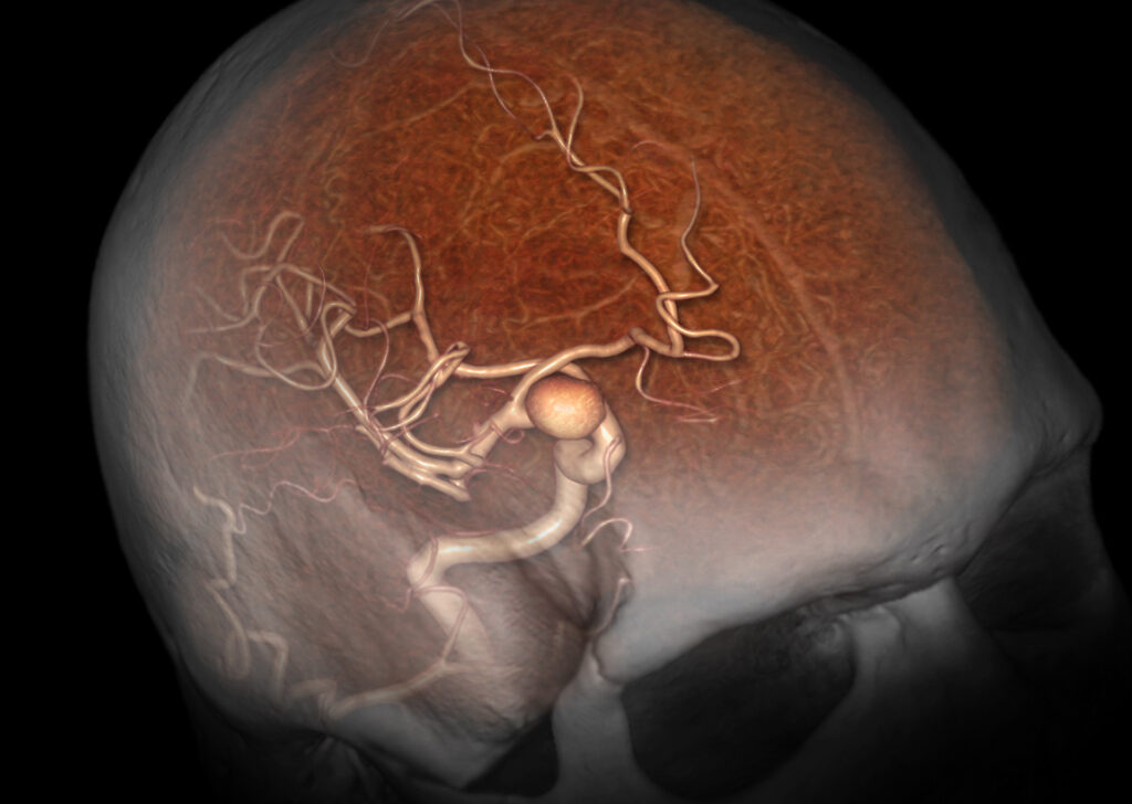
XA 3D Angio
XA 3D-Angio provides visualization and analysis tools for rotational images acquired in angiography labs. 3D angiography provides enhanced 3D views of complex anatomy.
Gain value with Enterprise Imaging
Explore Canon Medical’s Enterprise Imaging – with solutions focused on delivering clinician efficiency, enhancing, multi-departmental communication and cost-savings.


